A last-resort approach for PIH, nevus of Ota and stubborn congenital pigment — diagnosis first, staged combination care, including cases others decline.
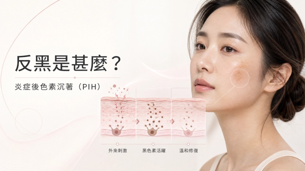
In medicine, “rebound darkening” is post-inflammatory hyperpigmentation (PIH): after external stimulus or injury, melanocytes become overactive and local skin tone deepens.
Common triggers include energy from light-based treatments, chemical peels, trauma or long sun exposure. When the skin’s defence is triggered, melanocytes produce more pigment to protect the area, forming uneven spots or patches.
PIH is a natural defensive response — not necessarily a medical error or failed treatment. If stimulus exceeds what the skin can tolerate, or pre- and post-care is inadequate, darkening may be stronger and last longer.
We focus on these complex pigment disorders, using precise skin assessment and gentle repair strategies to help the skin gradually clear excess pigment and restore even tone.
We use medical-grade devices including picosecond laser, fractional RF and related pigment technology. Devices do not cause PIH by themselves — incorrect parameters and poor judgement do.
Post-treatment pigment deposition is common in aesthetic medicine. From our clinical experience, frequent causes include:
These reflect operator training and experience, not the device itself.
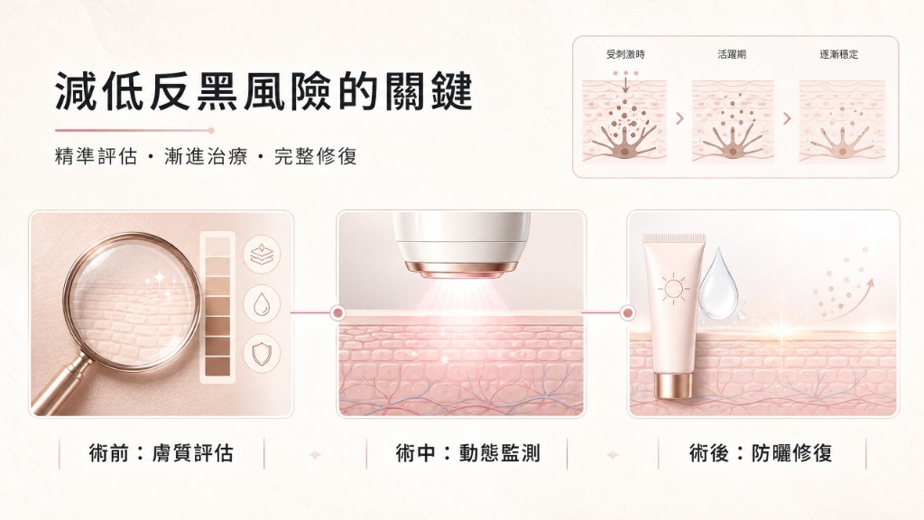
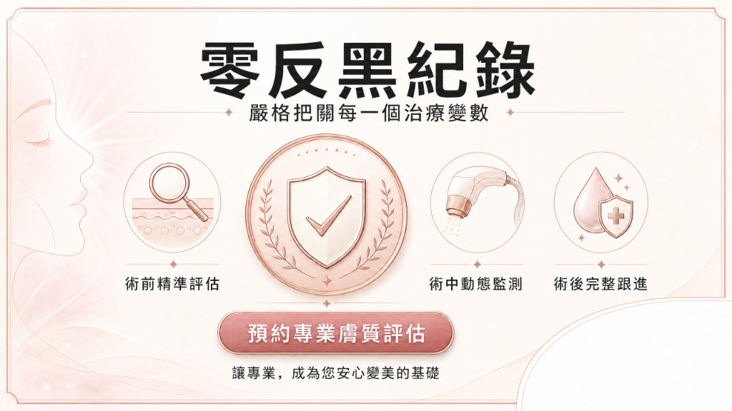
Our principle: control every variable — pre-treatment skin testing, device calibration, parameter tuning, in-treatment monitoring, and post-treatment follow-up — all to medical-grade standards.
Before — Detailed skin assessment and history; barrier status, melanin activity and prior treatments guide personalised parameters.
During — Low energy, step-by-step delivery; monitor response and keep heat accumulation within a safe range.
After — Full repair guidance: medical-grade skincare, sun protection schedule and review visits to support pigment clearance.
This systematic approach has helped many clients referred from other clinics with pigment problems. In our own treatments we have not seen post-procedure PIH — not by luck, but through years of clinical judgement and attention to detail.
Professionalism is not how many devices you own, but how precisely you use each one.
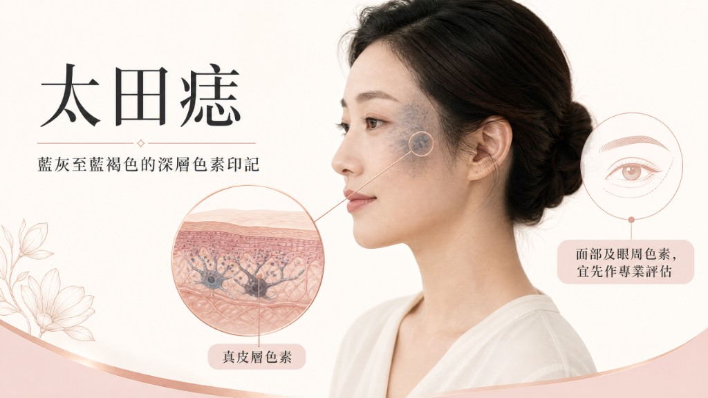
Nevus of Ota is a congenital dermal melanocytic hamartoma. Melanocytes abnormally cluster in the mid-to-deep dermis, forming grey-blue or brown-blue patches, usually in trigeminal distribution, present from birth or early childhood.
Pigment lies in the dermis, not only the epidermis; colour ranges from light grey-blue to deep brown-black, often with diffuse borders, and may deepen with age, sun or hormones.
It is a developmental anomaly, not sun-induced surface pigment. Superficial lasers often fail; incomplete improvement, recurrence or extra injury from wrong modalities has made it seem untreatable.
We treat these deep congenital lesions with pathology-led planning and dermal-targeted strategies to break down and clear ectopic melanocytes for stable, predictable improvement.
We use long-wavelength pigment lasers and Q-switched platforms. Devices alone do not remove nevus of Ota — depth assessment and wavelength/pulse choice determine success.
Poor outcomes often trace to:
Again, this reflects operator knowledge, not device failure.
We control every variable from imaging and depth mapping through calibration, in-treatment monitoring and aftercare — including strict sun protection and review to adjust the course and limit rebound pigment.
Many patients with prior poor results have seen meaningful improvement under this approach. Professionalism means reading the pathology and using each device with precision.
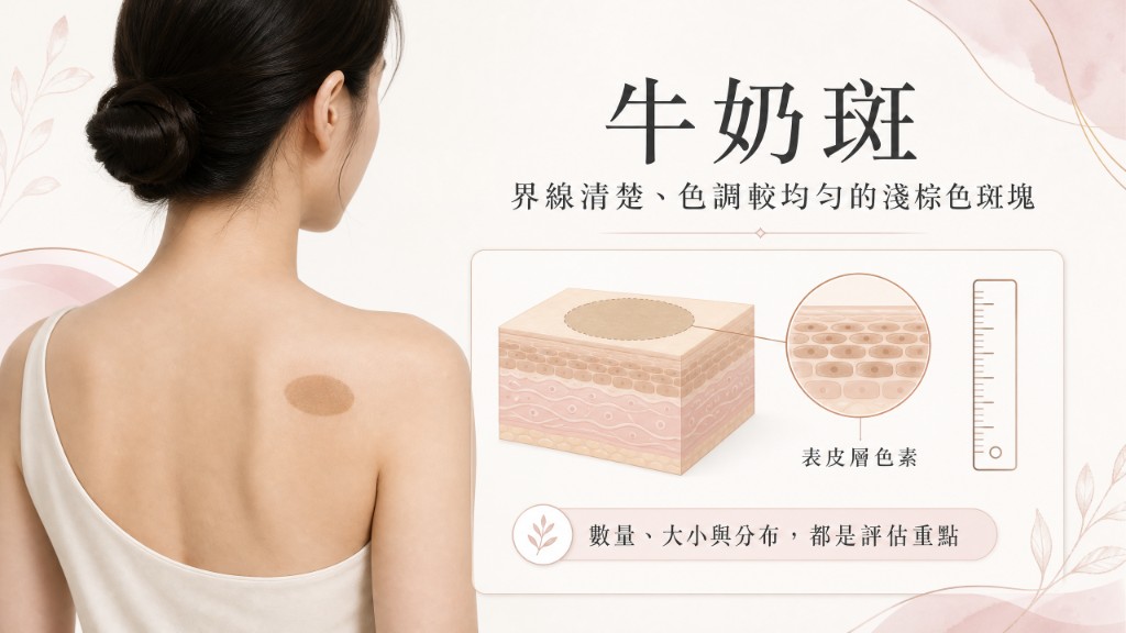
Café-au-lait macules are congenital superficial pigment lesions in the epidermis, light to deep brown with fairly clear borders, often visible from birth or childhood.
Pathology is increased melanocytes and melanin in the basal layer, usually not invading the dermis. Six or more lesions over 1.5 cm may suggest systemic conditions such as neurofibromatosis — differential diagnosis before treatment is essential.
Unlike nevus of Ota (deep dermal), milk spots are epidermal: treatment must avoid excessive surface injury and PIH, while Ota requires sufficient depth to reach dermal melanocytes.
Milk spots are not sun spots. Too little energy fades poorly; too much damages the epidermis and deepens colour through PIH.
We treat epidermal congenital pigment with typed assessment and epidermal-focused, gradual protocols.
We use Q-switched and picosecond pigment devices. Success depends on wavelength, pulse and energy matched to the lesion — not the brand on the label.
Common reasons for poor café-au-lait results include wrong wavelength, excessive energy, fixed protocols ignoring individual variation, poor intervals, and treating appearance without ruling out systemic disease.
Our workflow: imaging and history, calibration, low-energy staged treatment, selective photothermolysis with controlled heat, and structured aftercare with sun protection and follow-up.
Over years this has helped many café-au-lait patients achieve visible fading. Professionalism is pathology-first use of the right tool — that is why clients choose us.
PIH follows overstimulation of melanocytes after energy treatments, peels, injury or sun. We focus on assessment, staged energy and full aftercare.
Pigment sits deep in the dermis; superficial lasers often fail. Depth-matched wavelength, pulse settings and cooling are essential.
Epidermal café-au-lait spots can fade gradually with careful energy; multiple large lesions need systemic work-up first.
Skin imaging, history, pigment depth/activity, risk and expectations — then personalised parameters, not a sales pitch.